Creating an Interactive View of the Brain
Project Title: Virtual Reality System for Analyzing Human Brain Neuronal Networks
Team 17061 Members:
Erica Michelle Bosset, optical engineering
Joseph Elliott Clark, systems engineering
John Maximillian DiBaise, biomedical engineering
Josiah Michael McClanahan, electrical and computer engineering
Edward Richter, electrical engineering
Vincent Tso, biomedical engineering
Sponsor: UA Department of Biomedical Engineering
Project Gives Educators, Doctors New Tools

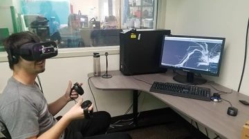
A team member wearing VR goggles tests out the system
In the iSpace of the University of Arizona Science-Engineering Library, five Engineering Design Program students on a 2018 senior design team take turns donning a virtual reality, or VR, headset and “entering” a room with a gray ceiling and gray walls. Directly in front of them is a shelf with two “brains” on it. Using VR hand controllers, the students pick up the brains from the shelf and examine them from every angle.
One brain is from magnetic resonance imaging. It looks like a scan of the swirly, dual-hemisphere brain with which most people are familiar. The other — a long, spindly representation of the brain’s white matter fiber tracts — is from a diffusion-tensor imaging scan.
Members of the interdisciplinary design team are fine-tuning their computer program to convert 2-D brain scans into 3-D images for viewing in a VR space.
The students, some working in a field outside their majors, say the project has given them a chance to contribute to a rapidly expanding area of technology.
“It’s a pure open-source, make-the-world-a-better-place, play-with-VR project,” said student team leader Joseph Elliott Clark, a systems engineering major. “What’s not to love?”
Benefits of 3-D Brain Images
When researchers and doctors look at a brain scan, they’re evaluating a 3-D object in a 2-D picture, and a lot of information gets lost in translation or becomes difficult to see. Similarly, it is difficult for students to learn about the brain’s complex structure from pictures, drawings, scans and other 2-D representations.
“Usually if we try to learn about the anatomy of the brain, we look at it the from angle one, angle two, angle three,” said Nan-Kuei Chen, UA associate professor of biomedical engineering, who is partnering with the program on the capstone project. “With this interactive mode, it’s not looking at only three angles — it’s looking at all possible angles.”
The senior student project, which will be on public display April 30 at the College of Engineering’s 2018 Design Day, will give researchers access to pre-uploaded brain images, including some that show symptoms of diseases such as Parkinson’s and Alzheimer’s. Ultimately, doctors diagnosing and treating brain conditions will be able to upload brain scans of their own patients for 3-D VR viewing.
Educational Add-ons
An added feature for education is audio that explains the functions of each anatomical region of the brain. For example, a user could use hand controls to select the medulla oblongata, and a voice might say, “Vital regions in the medulla oblongata regulate heartbeat, breathing, blood pressure, vomiting and coughing.” Another project extra, which Chen envisions as a free resource, is a VR Android app to help teachers explain basic brain anatomy to students.
“This is a very strong multidisciplinary team, and usually you don’t find so many talents all in the same group,” he said.

