Working Toward a Cure for Macular Degeneration

Project Title: Macular Degeneration Evaluation System
Team 17062 Members:
Erika Ackerman, biosystems engineering
Lexa Brossart, biomedical engineering
Shelley Meyer, biomedical engineering
Rory Morrison-Colvin, biomedical engineering
Ryan Nolcheff, optical sciences
Sponsor: UA Department of Biomedical Engineering
Students Develop Project That Could Help People See More Clearly
Macular degeneration is the leading cause of vision loss for people age 50 and over, and it affects approximately a third of people over 75.
A team of students in the University of Arizona’s Engineering Design Program are working on a device to study the disease’s causes by examining retinal pigment epithelial cells, or RPE. Their project could be a major step toward finding the first-ever treatment for macular degeneration.
Robert Snyder, an ophthalmologist and professor of biomedical engineering at UA, and Brian McKay, associate professor of ophthalmology in the UA College of Medicine, are the College project mentors.
McKay studies l-dopa, a type of protein that is produced by healthy RPE in a feedback loop in which cells produce the protein and the protein helps the cells grow. But in individuals with macular degeneration, l-dopa production has gone amok. McKay and Snyder also hypothesize that the proteins in the eye, including l-dopa, are produced in a sort of circadian rhythm, but that the rhythm is off in individuals with macular degeneration.
Putting a Hypothesis to the Test
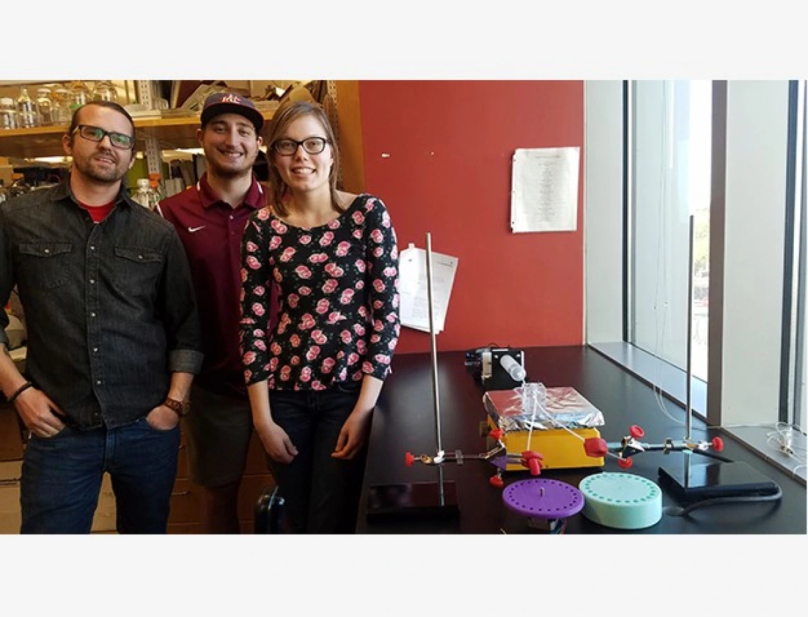
To test the hypothesis, the students are creating an apparatus designed to mimic the environment of the eye and test levels of protein output throughout the day.
Nutrients such as l-dopa move via two tubes through a chamber of RPE, and come out the other end of the chamber carrying new proteins produced by the RPE. The system is unusual in that it washes the nutrients over the cells rather than leaving cells in a static pool of media.
“If you had a culture system that was just a bunch of cells lying in a culture plate for 24 or 48 hours, you wouldn’t see that feedback loop,” Snyder said.
RPE cells are polarized, with apical surfaces that face the external environment and basal surfaces that face the internal environment. The two-tube system corresponds with the two polarities and will allow researchers to see which proteins are being produced by which surfaces.
The nutrients with the new RPE proteins will be collected in microcentrifuge dishes on two collection disks, with 24 dishes each that rotate every hour. This means there will be a sample of both the apical and basal proteins produced for every hour of the day. An enzyme-linked immunosorbent assay machine, or an ELISA, can then determine the protein levels in the samples.
“Essentially, we’ll have a time-dependent scale of each of the proteins throughout the day,” said student team leader Erika Ackerman.
Once the system — which will be on public display at the College of Engineering’s 2018 Design Day on April 30 — is up and running, the team and future researchers can experiment with variables. Such variables could include exposing the RPE to 12 varying periods of darkness and light, or adjusting the nutrients and flow rate to investigate other RPE-related diseases such as albinism and glaucoma.

